SCLERA & CONJUNCTIVA
EPISCLERITIS
DEF-
Benign inflamn of subconj connective tiss & superficial scleral lamella.
Young adults
Females
H/O RA
TYPES- Simple
-Nodular
C/F-
Symptoms-UL
-Acute redness
-discomfort
-tenderness
-watering
Signs-
1.SIMPLE EPISCLERITIS-
a] Sectoral OR
B] Diffuse
* D/T engorgement of large episcleral Vs running in a radial dir beneath the conj
* Never ulcerate
* Cor & uvea NOT inv
* May resolve or leave a slate coloured scar to which conj is adherent
2.NODULAR EPISCLERITIS-
* Localized, raised , congested nodule
* Ant scleral surf is not raised
* Traversed by deeper episcleral Vs.So look purple & not red.
T/T-1. Lubricants
2.Topical steroids
3. NSAIDs- T. Flurbiprofen 100mg tds
- OR t. Aspirin
- T. Indomethacin
SCLERITIS [j-01, d-02, j-04]
DEF-
Edema & cellular infiltration of entire scleral thickness.
CAUSES-
1.Systemic-
* RA
* Wegener’s granulomatosis
* Relapsing polychondritis
* PAN
2.Surgery-
* Cataract surg
* RD surg
* Filtration surg
* pterygium excision + beta rad + MMC
3.Infections-
* Corneal ulcer
*Trauma
* Org- Pseudo aeruginosa, strep pneum, staph aureus, varicella zoster
CLASS-
Anterior scleritis-
Non-necrotizing-Diffuse or nodular
Necrotizing- With or without inflamn
2.Posterior scleritis
ANTERIOR NON-NECROTIZING SCLERITIS-
1 ]Diffuse scleritis-
* Inv a sector or entire anterior sclera
* Distorts the nml radial vascular pattern
2] Nodular scleritis-
* Scleral nodule cannot be moved over the underlying tiss
* Dark red or bluish, later purple
T/T-
1.Oral NSAIDs
2.Oral steroids
3. Steroids + NSAIDs
4.S/C Triamcinolone acetonide 40mg/ml
ANTERIOR NECROTIZING SCLERITIS-
WITH INFLAMN-
Localized redness
Pain-radiates to temple,brow or jaw
Congestion of deep vascular plexus.
Vascular distortion & occlusion → avascular patches
Scleral necrosis with overlying conj ulceration
Resolution → scleral thinning→uvea visible as bluish tinge
COMPLICATIONS-Staphyloma
-Cataract
-Keratitis
-Keratolysis
-Sec glaucoma
-Macular edema
T/T-
1.Oral steroids
2.Immunosuppressives-
Cyclophosphamide-1-2 mg/kg/day
Azathioprine-1-3mg/kg/day
Cyclosporin-2-5 mg/kg/day
3.Combined therapy-
Pulsed IV methylprednisone 1gm + cyclophosphamide 500mg
ANTERIOR NECROTIZING SCLERITIS
WITHOUT INFLAMN-
Also k/as –SCLEROMALACIA PERFORANS
Asymptomatic yellow ,necrotic,scleral patches in uninflamed sclera
Scleral thinning→ exposes uvea
Staphyloma
T/T- ineffective
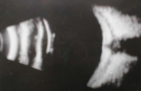
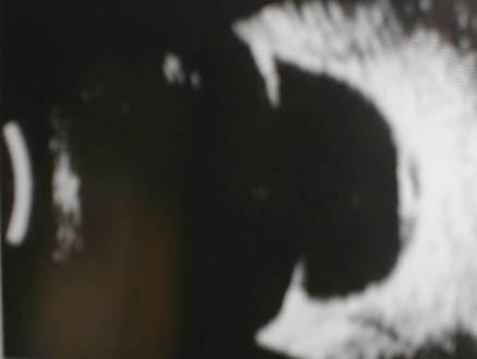
POSTERIOR SCLERITIS-
C/F-
Pain
Visual impairment
Lid edema
Proptosis
O’plegia
Asstd ant scleritis
Fundus-Disc edema, mac edema,choroidal folds,exud RD,Ring choroidal det& subretinal lipid exud
INV-
USG-“ T” sign-Thickening in post sclera & fld in tenon’s space.Stem of T is formed by optic N & cross-bar by fld in subtenon’s spa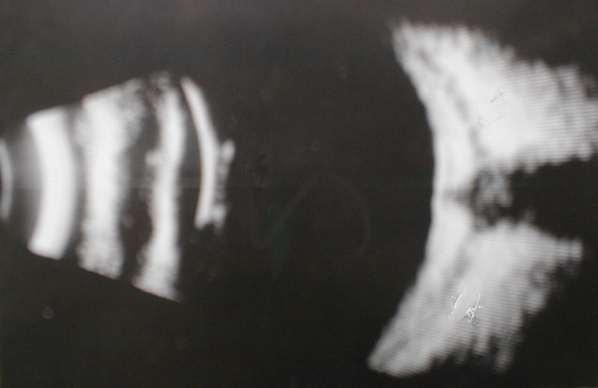 ce
ce
CT SCAN- Posterior scleral thickening
T/T- same
STAPHYLOMA-case
DEF-
Ectasia of outer coats of the eye with an incarceration of uveal tissue
Underlying cause is inflammation / degeneration
CLASS-
Anterior
Intercalary
Ciliary
Equatorial
Posterior
ANTERIOR STAPHYLOMA-
Partial- Part of cornea inv
Total-Whole of cornea inv
MCC- Sloughing corneal ulcer→ perforates → Heals & forms pseudocor by exudative organization & laying down of fibrous tissue
Lined internally by iris & externally by epith
AC-flat
Sec glaucoma dev later
Gradually weak ant surf of eye protrudes out→ Ant staphyloma
INTERCALARY STAPHYLOMA-
Perforating inj of peripheral cor
Marginal cor ulcer
Ant scleritis
Scleromalacia perforans
Complicated cataract surg with poor wound apposition
Sec glauc
3.CILIARY STAPHYLOMA-
* Affects the ciliary zone [8mm behind the limbus]
* CB is incarcerated in scleral ectasia
* Bluish & lobulated
Causes-
1.Dev glauc
2.End stage glauc
3.Scleritis
4.Trauma to ciliary region
4.EQUATORIAL STAPHYLOMA-
* Occurs at the equatorial reg with incarceration of choroid [14mm behind the limbus]
* Equator is Weak d/t passage of venae vorticosae
Causes-
Scleritis
deg myopia
Chr uncontrolled glauc
POSTERIOR STAPHYLOMA-
Posterior pole of eye
Lined by choroid
MCC- deg high axial myopia
Ectatic portion detected externally
Fundus-Crescentic shadow in macular region
-Retinal Vs change dir dipping into the region
*Staphylomatous area- pale d/t deg in ret, RPE &
Choroid
T/T-
T/T of underlying cause
Local excision & repair with corneal & scleral patch graft
Unsightly & blind eye-Staphylectomy & KP
OR Enucleation + implant
PARTS OF CONJUNCTIVA-
1. Palpebral-
Marginal
Tarsal
Orbital
2.Fornix
3.Bulbar
4.Limbal
MICROSCOPIC-
EPITHELIUM
Palpebral conj- 2 layers of epith
Intermarginal strip-Transitional stratified squamous epith
From fornix to limbus-4-6 layers
Limbus- stratified epith
SUB-EPITHELIAL / ADENOID LAYER-
Loose connective tiss + leucocytes
C . FIBROUS LAYER
XEROPHTHALMIA-WHO CLASS-
XN- Nightblindness
XIA- Conjunctival xerosis
XIB- Bitot’s spots
X2- Corneal xerosis
X3A- Corneal ulceration/keratomalacia <1/3rd cor surf
X3B- Corneal ulceration / keratomalacia > 1/3rd cor surf
XS- Corneal scar
XF- Xerophthalmic fundus [UYEMURA’S FUNDUS]
Night blindness is also k/as –Chicken eyes [since chickens lack rods & are nightblind]
CONJUNCTIVAL CONGESTION
|
CILIARY CONGESTION
|
Bright red
|
Dull red
|
Near the fornix
|
Around the limbus
|
Branch dichotomously
|
Branch radially
|
Arise from post conj Vs
|
Arise from anterior ciliary Vs
|
Phenylepherine→ blanch
|
Do not blanch
|
Vs fill up from the fornix
|
Vs fill up from the limbus
|
Superficial inv-c’vitis, simple hyperaemia
|
Deep inv-iritis, scleritis
|
TRUE PTERYGIUM
|
PSEUDOPTERYGIUM
|
Degenerative
|
Inflammatory
|
Usually progressive
|
Stationary
|
Probe cannot be passed underneath the head of pterygium
|
Probe can be passed.
|
FOLLICLES-
Localised aggregation of lymphocytes in the subepithelial adenoid layer.
CAUSES-
HSV conjunctivitis
molluscum contagiosum c’vitis
chlamydial inf
Parinaud’s oculoglandular syn
Q Diff bet trachomatous folliclec & follicles in follicular conjunctivitis?
A- Trachomatous follicles- 5mm dia
-Commence in lower fornix
-Form a row along upper tarsal margin
-Undergo cicatrisation & form minute
Stellate scars.
PAPILLAE-
Hyperplasia of central Vs surrounded by diffuse infiltrates of lymphocytes,plasma cells & eosinophils
CAUSES-
Chr blepharitis
Vernal catarrh
Giant papillary c’vitis
contact lens induced
Superior limbic keratoconjunctivitis
FOLLICULAR CONJUNCTIVITIS [d-04]
1.Acute
2.Subacute / chronic
ACUTE FOLLICULAR CVITIS-
Chlamydial inclusion conjunctivitis
Epidemic keratoconjunctivitis
Pharyngoconj fever
Newcastle cvitis
H’hic cvitis
Primary herpetic cvitis
Recurrent herpes simplex cvitis
SUBACUTE /CHRONIC
1) CHLAMYDIAL INCLUSION CVITIS-
Agent-Chlamydia trachomatis[D-K]
Spread-genitals,eye-eye,swimming pool
C/F- UL/BL mucopurulent discharge
SIGNS-large follicles in lower fornix→2-3wks→SPK + pannus
T/T-
Topical tetracycline e/o qid for 6wks
Tab doxy 100mg 12hrly for 2wks
Tab erythro 250mg 12 hrly for 2wks
Tab. Azithro 1gm OD
2) EPIDEMIC KERATOCONJUNCTIVITIS-
-Preauricular LNpathy
-Punctate epith infiltrates
-discrete subepith opacities
* T/T- Decongestive & lubricating drops
-Antibiotic e/d
3) PHARYNGOCONJUNCTIVAL FEVER-
Agent-adenovirus3,4&7
C/F-
Foll cv
Pharyngitis
Fever
Preaur LNpathy
SPK
4) NEWCASTLE CV
5) H’GHIC CV
6) ACUTE HERPETIC CV
7) RECURRENT HERPES SIMPLEX CV
Artificial tears
acyclovir e/o 3% 5 /day
Vidarabine e/o 3% 5/d
Trifluorothymidine 1 % e/d 5/d
VERNAL CATARRH [j-04, d-02]
-Sporadic
-non-contagious
-BL
-Hot weather
-Young boys
-F/H of atopy
-Type –I hypersensitivity-Ig E mediated
-eosinophilia
* C/F-
-Burning
-Itching
-Photophobia
-Lacrimation
-White ropy discharge [b/o fibrin]
2 forms-1] Palpebral
2] Limbal / Bulbar
PALPEBRAL-
Palp conj is hypertrophied [papillae]& mapped out into polygonal raised areas like cobblestones
Bluish –white like milk
Flat topped nodules are hard & consist of dense fibrous tiss.The overlying epith is thickened,therefore milky hue.
BULBAR-
COMPLICATIONS-
SPK
Dry eyes
Corneal [ shield] ulcer
Scarring
T/T-
Cold compression & tinted glasses
Antihistaminic e/d
Topical steroid e/d 4-6 hrly
Disodium chromoglycate e/d 2% 8 hrly or 4% 12 hrly
Olopatadine e/d BD- new mast cell stabilizer
Subtarsal inj of triamcinolone
Acetyl cysteine 10 % or 20 % drops-for excess mucus
Cryotherapy of nodules
ANTI-ALLERGIC DRUGS [j-07]
H1- RECEPTOR ANTAGONIST [ANTIHISTAMINICS]
Competetively inhibit histamine at the receptor sites
IND-
VKC
GPC
Allergic cvitis
TOPICAL-
Emedastine qid
Levocabastine
Azelastine OD or BD
Antazoline qid
Chlorpheniramine qid
SYSTEMIC-
Loratidine
Cetrizine
Astemizole
Fexofenadine
MAST CELL STABILIZERS-
Stabilize the membrane of mast cells→ prevent release of histamine
Cromolyn sodium-
-2-4 % E/D 6 hrly
- 2 % oint HS
- Ind- VKC
-GPC
- onset of action- 3-4 weeks
Ketotifen –
TDS
Quicker onset of action
Lodoxamide-
- 0.1 % TDS
Olopatadine-
Others- pemirolast , nedocromil sodium
3] ANTIHISTAMINES + MAST CELL STABILIZERS-
* Olopatadine
* Ketotifen [ anti-inflam]
* Azelastine [ “ ]
4] NSAIDs
- Ketorolac- Reduces itching
- but stinging
5] VASOCONSTRICTORS-
-Naphazoline / pheniramine
- Naphazoline / Antazoline
6] STEROIDS-topical
- Loteprednol
- Flurometholone
- Rimexolone