Ophthalmology Notes @ OphthalNotes.blogspot.com

Ophthalmology Notes @ OphthalNotes.blogspot.com
A comprehensive collection of ophthalmology revision notes that cover a broad range of topics.
Showing posts with label EASY TO REMEMBER. Show all posts

Rings in ophthalmology

Rings in ophthalmology

1-Ring Keratitis: The hallmark of Acanthamoeba keratitis.


2-Kayser-Fleischer’s ring: Wilson’s disease


3-Corneal rust ring: 

A small, reddish brown, circular opacity remained in the cornea after the removal of an iron foreign body.


4-Coats’ white ring: 

Remnants of a foreign body. The remnants are fine iron deposits in the cornea.


5-Fleischer’s ring: 


Visible all around the base of cone in Keratoconus

6-Pseudo-Fleischer’s ring: 

Iron deposition can be seen in Hyperopia LASIK correction

7-Soemmering’s ring: 

Early opacification of lens capsule after cataract extraction

8-Vossius’ ring: 


Iris Pigment on anterior lens capsule in concussion injury to eye

9-Weiss ring: 


Epipapillary glial tissue torn from the optic disc in Posterior vitreous detachment (PVD)

10-Double ring sign: 


With the peripheral margin of the encircling ring corresponding to the border of a normal-sized optic disc. Seen in Hypoplasia of the Optic Disc.

11- Golden Ring : 


A golden ring within the lens is evidence of successful delineation.

12-Intrastromal Corneal Ring Segments (ICRS):

are implanted in the deep corneal stroma to modify the corneal curvature. Used in keratoconus,PMD. 

13-Capsular tension rings (CTRs) :

were used for zonular reinforcement in eyes with a weak zonular apparatus, such as in pseudoexfoliation and Marfan's syndrome, and when zonular rupture or dehiscence occurs after blunt or surgical trauma.

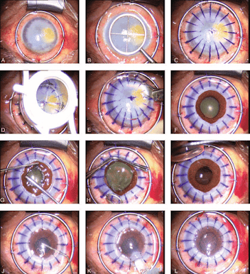
14-Flieringa ring: 


A stainless steel ring sutured to the sclera to support the globe during difficult eye operations. used usually in corneal transplant surgeries 

15-Wessely immune ring: 

refers to the formation of a ring-shaped infiltrate in the corneal stroma that arises from a type 3 immune response involving antigen-antibody complex formation.

SPOTS & DOTS in Ophthalmology


SPOTS & DOTS in Ophthalmology

1. Cherry red spot – 

Red colour of fovea contrast to the milky retinal odema seen in CRAO

2. Foster Fuchs spot - 

Myopic retinopathy (resolved haemorrhage & pigment)

3. Roth’s spot - 

Retinal haemorrhage with clear centre composed of coagulated fibrin including, inflammatory infiltrates, infectious organisms or neoplastic cells in SABE, anemia, leukemia

4. Elschnig spot- 

Yellow isolated area in retinal choriocapillary occlusion (choroidal infarcts)

5. Bitot’s spot - 

Keratinised superficial conjunctival spots in vitamin A deficiency (late stage)

6. Brush field spot - 
White or greyish brown spots on periphery of iris in Down syndrome

7. Fischer-Khunt spot - Senile scleral paque, area of hyalinised sclera anterior horizontal rectus muscle insertion, seen in old age

8. Gunn’s spot - 
Light reflections from internal limiting membrane around disc and macula

9. Kaye's spot - 
White punctate epithelial opacities anterior to suture line of corneal graft

10. Mittendorf spot - 
Anterior remnant of hyaloid artery at posterior surface of lens

11. Map dot fingerprint dystrophy- 
Corneal epithelial basement membrane dystrophy (multilamellar basement membrane)


Rings in ophthalmology

1-Ring Keratitis: The hallmark of Acanthamoeba keratitis.


2-Kayser-Fleischer’s ring: Wilson’s disease


3-Corneal rust ring: 

A small, reddish brown, circular opacity remained in the cornea after the removal of an iron foreign body.


4-Coats’ white ring: 

Remnants of a foreign body. The remnants are fine iron deposits in the cornea.


5-Fleischer’s ring: 


Visible all around the base of cone in Keratoconus

6-Pseudo-Fleischer’s ring: 

Iron deposition can be seen in Hyperopia LASIK correction

7-Soemmering’s ring: 

Early opacification of lens capsule after cataract extraction

8-Vossius’ ring: 


Iris Pigment on anterior lens capsule in concussion injury to eye

9-Weiss ring: 


Epipapillary glial tissue torn from the optic disc in Posterior vitreous detachment (PVD)

10-Double ring sign: 


With the peripheral margin of the encircling ring corresponding to the border of a normal-sized optic disc. Seen in Hypoplasia of the Optic Disc.

11- Golden Ring : 


A golden ring within the lens is evidence of successful delineation.

12-Intrastromal Corneal Ring Segments (ICRS):

are implanted in the deep corneal stroma to modify the corneal curvature. Used in keratoconus,PMD. 

13-Capsular tension rings (CTRs) :

were used for zonular reinforcement in eyes with a weak zonular apparatus, such as in pseudoexfoliation and Marfan's syndrome, and when zonular rupture or dehiscence occurs after blunt or surgical trauma.

14-Flieringa ring: 


A stainless steel ring sutured to the sclera to support the globe during difficult eye operations. used usually in corneal transplant surgeries 

15-Wessely immune ring: 

refers to the formation of a ring-shaped infiltrate in the corneal stroma that arises from a type 3 immune response involving antigen-antibody complex formation.

PHENOMENON in Ophthalmology

PHENOMENON in Ophthalmology

1. A&V phenomenon 

Deviation of eyeball on upgaze and downgaze


2. Bells phenomenon  

Upward rolling of eyeball on lid closure


3. Entoptic phenomenon 


Visualisation of one’s own ocular structure


4. Fish egg phenomenon  


Silicon oil dispersion in replaced vitreous

5. Haidinger phenomenon  - Seeing  radial pattern of  macula on entoptic visualisation.


6.  Jaw winking phenomenon 

Lid retraction on movement of jaw (aberrant regeneration)


7.  Loch ness  phenomenon

Loop of vessel  on gonioscopy of infant


8. Pulfrich’s phenomenon 

Perception defect of movement in two eyes due to differential signal timings in optic neuritis.


9.  Pseudo Graefe phenomenon - 

Ptosis improves on adduction in 3rd nerve palsy (aberrant regeneration)

10.  Riddoch’s phenomenon - 

Perception of movement not the spot(cortical lesion) 
Riddoch syndrome (also known as the Riddoch phenomenon) is an ocular affectation often caused by lesions in the occipital lobe which limit the sufferer's ability to distinguish objects. Only moving objects in a blind field are visible, static ones being invisible to the patient. The moving objects are not perceived to have color or detail. 

The subject may only have awareness of the movement without visual perception of it (gnosanopsia), or the general shape of a moving object may be perceivable as a shadow like outline.
 

11.  Uthoff ’s phenomenon - 

Decreased visual acuity on exertion (heat) in optic neuritis (multiple sclerosis)