Ophthalmology Notes @ OphthalNotes.blogspot.com

Ophthalmology Notes @ OphthalNotes.blogspot.com
A comprehensive collection of ophthalmology revision notes that cover a broad range of topics.
Showing posts with label EASY TO REMEMBER. Show all posts

Triads & Syndromes in Ophthalmology

Triads & Syndromes in Ophthalmology

Triads are usually a combination of three clinical features or signs, often used to describe various clinical conditions or diseases. Here we have compiled some of these triads used in ophthalmology.

Bálint's Syndrome= Simultanagnosia+ Optic ataxia+ Ocular motor apraxia

Behçet's Disease= Oral ulcers + Genital ulcers + Hypopyon uveitis

Cerebral Whipple's Disease= Somnolence + Dementia + Ophthalmoplegia.

Cone Degeneration= Progressive central acuity loss + Color vision disturbances + Photophobia.

Congenital Glaucoma= Epiphora + Blepharospasm+ Photophobia

Congenital Rubella Retinopathy= Cataracts+ Deafness + Congenital heart disease

Congenital Toxoplasmosis= Retinochoroiditis+ Hydrocephalus+ Intracranial calcifications

De Morsier's Syndrome= Short stature+ Nystagmus + Optic disc hypoplasia

Fechtner's Syndrome= Nephritis+ Sensorineural hearing loss+ Eye abnormalities

Gaucher Disease= Trismus+ Strabismus+ Opisthotonus

Horner's Syndrome= Ptosis+ Miosis + Ipsilateral anhidrosis of the face

Intraoperative Floppy Iris Syndrome=A flaccid iris stroma that undulates and billows in response to ordinary intraocular fluid currents+ A propensity for the floppy iris stroma to prolapse toward the phaco and side-port incisions, despite proper wound construction + Progressive intraoperative pupil constriction despite standard preoperative pharmacologic measures designed to maximize dilation

Kearns–Sayre Syndrome=External ophthalmoplegia + Pigmentary retinopathy + Cardiac conduction block during the first or second decade of life

Lambert–Eaton Myasthenic Syndrome=Muscle weakness+ Autonomic dysfunction+ Hyporeflexia

Miller-Fisher Syndrome=Ataxia+ Ophthalmoplegia+ Areflexia

Ocular Ischemic Syndrome=Midperipheral dot hemorrhages+ Dilated retinal veins + Iris neovascularization

Ocular Tilt Reaction=Skew deviation+ Cyclotorsion of both eyes + Paradoxical head tilt

Optic Nerve Sheath Meningioma=Optociliary venous shunts on the disc+ Diffuse disc edema (eventually replaced slowly by pallor)+ Insidious visual loss

Osteogenesis Imperfecta=Brittle bones+ Blue sclera+ Deafness (otosclerosis)

Pharyngoconjunctival Fever=Fever+ Pharyngitis + Acute follicular conjunctivitis

Pierre Robin Syndrome=Micrognathia+ Glossoptosis+ Cleft palate

Pigmentary Glaucoma=Corneal pigmentation (Krukenberg's spindle)+ Slit-like, radial, mid-peripheral iris transillumination defects+ Heavy accumulation of pigment in the trabecular meshwork

Presumed Ocular Histoplasmosis Syndrome=Peripapillary atrophy+ “Punched-out” chorioretinal lesions + Disciform macular scarring in young and middle-aged adults

Reiter's Syndrome=Arthritis+ Urethritis+ Conjunctivitis

Schwartz's Syndrome=Rhegmatogenous retinal detachment+ Uveitis+ Glaucoma

Sjögren's Syndrome=dry eyes+ dry mouth+ Arthritis (dry joints)

Spasmus Nutans=pendular nystagmus+ head nodding+ torticollis

Sturge-Weber Syndrome=Port wine facial telangiectasis (nevus flammeus) in the distribution of the trigeminal nerve that respects the vertical midline+ Ipsilateral glaucoma (ipsilateral buphthalmos)+ Contralateral seizures caused by ipsilateral leptomeningeal hemangiomatosis

UGH Syndrome=Uveitis+ Glaucoma + Hyphema

Source:EOphtha

Lines in Ophthalmology

 Lines in Ophthalmology

1.Arlt’s Line 

= conjunctival scar in sulcus subtarsalis in Trachoma.

Horizontal line of conjunctival scarring in sulcus subtarsalis of superior palpebral conjunctiva.

2.Ehrlich-Turck Line

 = linear deposition of KPs in uveitis

3.Ferry’s Line 

= corneal epithelial iron line at the edge of filtering blebs.

4.Hudson-Stahil Line

= Horizontal corneal epithelial iron line at the inferior one third of cornea due to aging.
Iron deposition line in the corneal epithelium, which commonly seen in the junction between middle and lower third cornea (upper border of normal tear layer strip).
Often seen in the elderly.
Causes no symptoms or clinical significance.

5.Khodadoust Line 

= corneal graft endothelial rejection line composed of inflammatory cells.

Classic endothelial rejection presents with an endothelial rejection line (Khodadoust line) that usually begins at a vascularized portion of the peripheral graft-host junction and progresses, if untreated, across the endothelial surface over several days. The rejection line consists of mononuclear white cells that damage endothelial cells as the line sweeps across the endothelium.

6.Paton’s Line 

= Circumferential retinal folds due to optic nerve edema.

7.Sampaoelesi line


= Increased pigmentation anterior to Schwalbe’s line in pseudoexfoliation syndrome.

Occurs in

  • pseudoexfoliation syndrome
  • pigment dispersion syndrome
  • trauma
  • iris melanoma

8.Zentmeyer line (Scheie’s Line):

 Pigment on the equatorial surface of the lens and posterior capsule in pigment dispersion syndrome.

"Scheie's line" is considered to be pathognomonic for pigment dispersion syndrome.

9.Schwalbe’s Line 

= Angle structure representing peripheral edge of Descemets membrane.

10.Stockers Line 

= Corneal epithelial iron line at the edge of Pterygium

11.White lines of Vogt 

= Sheathed or sclerosed vessels seen in Lattice degeneration

12.Fingerprint lines: 

The map-dot fingerprint dystrophy

13.LASIK Iron Line: 

After LASIK for myopia, the central corneal curvature is flatter than before surgery. The tear film distribution is therefore altered, allowing some pooling centrally. This pooling can cause iron deposition in the central epithelium. A similar effect can be seen after steeping of the cornea from treatment of hyperopia. In the case of hyperopia, a pseudo-Fleischer’s ring iron deposition can be seen. These iron lines do not affect vision.

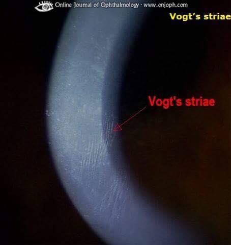
14.Vogt’s striae 

it occur centrally in a patient with keratoconus. By applying digital pressure on the eye while looking through the slit lamp, these striae in the deep cornea, mostly Descemet’s membrane, disappear, which is charac teristic of keratoconus.

15.The Descemet’s breaks or Haab’s striae 

The Descemet’s breaks or Haab’s striae from birth trauma tend to be vertical, while the Descemet’s tears associated with congenital glaucoma tend to be horizontal or curvilinear.


Eponyms and their characteristics

 Eponyms and their characteristics.

Eponym

Location

Description / context / pathophysiology

Relevance

Hudson-Stahli line

Cornea

Brown-green, horizontal line. Middle to inferior 1/3 cornea.

Significance: iron deposition, usually with age. Enhanced by chloroquine or hydroxychloroquine.

Not usually visually significant

Waring line

Cornea

Stellate corneal epithelial iron deposition. After radial keratotomy (RK).

Present in up to 80% of RK eyes.

Not usually visually significant

Khodadoust line

Cornea

Line on the corneal endothelium in an eye with a corneal graft. Keratoplasty / corneal graft surgery.

Mononuclear cells / keratic precipitates accumulating on the corneal endothelium.

Corneal graft endothelial cell rejection

Eponym

Location

Description / context / pathophysiology

Relevance

Paton’s lines

Retina / optic disc

Vertical, circumferential retinal folds especially temporal to the optic disc.

On direct ophthalmoscopy they are better visualised when the light on ophthalmoscope is moved back and forth.

Optic nerve head swelling, protrusion and eventual corrugation of the retina.

Blockage of the axoplasmic transport at the lamina cribrosa.

Optic disc swelling

Sampaolesi’s line

Drainage angles

Pigment deposition along Schwalbe’s line.

Pseudoexfoliation, pigment dispersion syndrome, iris melanoma, trauma

Scheie’s line

Lens

Also see Zentmayer ring.

Pigment accumulated at the zonular attachments to the lens. Although some report it as pigment on peripheral posterior lens capsule.

Pigment dispersion syndrome

Arlt’s line

Conjunctiva

Horizontal scarring of the upper tarsal conjunctiva at the junction of the anterior one third and posterior two thirds of the conjunctiva.

Typically seen in trachoma. Chronic inflammation of tarsal conjunctiva

Stocker’s line

Cornea

Yellow or brown deposition in epithelium.

Iron deposition can be seen adjacent to the leading edge of pterygium.

Pterygium

White lines of Vogt

Retina

Sheathed or sclerosed vessels in lattice degeneration.

Lattice degeneration

Vogt striae

Cornea

Vertical stromal / Descemet’s membrane lines. Stress lines due to stretching and thinning.

Disappear with globe pressure.

Keratoconus

Haab striae

Cornea

Horizontal or concentric breaks in Descemet’s membrane. Similar to posterior polymorphous dystrophy (PPMD). However on histopathology: the edge of Haab’s striae are thickened, curled, with the area between the edge being smooth and thin. This helps differentiate from PPMD.

Congenital glaucoma

Ohngren’s line

Orbit

An x-ray description from 1930.

Delineates the limits of resectability of maxillary sinus tumours. If superoposterior it is more likely to invade orbit, ethmoids and paterygopalatine fossa.

Its use is less certain due to a difference in surgical techniques and treatments

Ferry line

Cornea

Corneal epithelial line at the edge of trabeculectomy blebs. Iron deposition.

No visual significance

Ehrlich-Turck line

Cornea

Linear deposition of KPs on corneal endothelium. Uveitis.

Uveitis

Schwalbe’s line

Drainage angles

Clinical: gonioscopic view of the drainage angles. Delineates anterior edge of trabeculum and termination of Descemet’s membrane.

Recognition, last to angle structure to disappear in narrow angles

Siegrist streaks

Choroid

Hyper-pigmented flecks that are arranged in a linear fashion along the choroidal blood vessels.

Hypertensive choroidopathy. Fidrinoid necrosis.

Hypertension Giant cell arteritis

Linear naevus sebaceous of Jadassohn

Periorbit

A congenital hairless plaque that is usually found on the scalp, face or neck.

Schimmelpenning syndrome: triad of: sebaceous nevi, seizures and learning difficulties. Ocular: coloboma, choristomas, e.g. posterior scleral cartilage

Kayes dots

Cornea

Subepithelial infiltrates seen in corneal graft rejection. Could be an elevated line.

Corneal graft rejection

Gunn’s dots

Retina

These are visible reflections of the internal limiting membrane, created by the footplate of the Muller cells. Described in 1918. Note Marcus Gunn was using an ophthalmoscope based on a mirror and a solid flame as an illuminating source to find them.

Significance: Uncertain. Described as a cause for photosensitivity

Horner-Trantas dots

Cornea

Gelatinous dots at the corneal limbus. Chalky collections of eosinophils at limbus.

Vernal Keratoconjunctivitis

Mittendorf’s dot

Lens

Whitish spot (on direct illumination) at posterior lens surface. Black in retroillumination.

Usually nasal / inferonasal.

Embryological remnant of the hyaloid artery.

Associated posterior polar cataract, care as associated with posterior capsule

(PC) rupture risk during hydrodissection

Eponym

Location

Description / context / pathophysiology

Relevance

Bitot’s spot

Conjunctiva

White, foamy area (oval / triangular / irregular in shape) on conjunctiva.

Conju quamous metaplasia of bulbar conjunctiva with keratin layer.

Vitamin A deficiency Xerophthalmia

Elschnig spots

Choroid

Hyperpigmented patches in the choroid surrounded by a ring of hypopigmentation.

Choriocapillaris hypoperfusion.

Hypertension

Fuchs spot

Retina

Pigmented macular lesion.

Significance: retinal pigment epithelium (RPE) hyperplasia / degeneration.

Forster-Fuchs’ retinal spot: subretinal neovascularisation.

Pathological myopia

Brushfield spot

Iris

White, grey spots in peripheral iris.

Iris stromal hyperplasia and surrounding hypoplasia.

Down’s syndrome present in around 78%

Roth’s spots

Retina

Haemorrhages with white centre.

Platelet and fibrin thrombus at the centre of a ruptured capillary network.

Also: immune complex mediated vasculitis.

Numerous e.g.:

   Subacute bacterial endocarditis

   Leukemias

   Anaemia

   Anoxia

   Carbon monoxide poisoning

   Eye decompression

Koplik’s spot

Conjunctiva

Occur on the conjunctiva and resemble specks of sand surrounded by a red areola.

Also curuncle lesion and semilunar fold (Hirschberg’s sign).

Measles

Fischer-Khunt spot

Sclera

Blue grey plaque anterior to horizontal recti insertions. Senile scleral plaque.

Area of hyalinised sclera.

Seen in old age

Krachmer spots

Cornea

Sub-epithelial opacities similar in appearance to adenovirus keratitis.

Present Bowman’s layer.

Note: Stromal swelling can coincide leading to some calling it a stromal rejection.

Corneal graft epithelial rejection

Kayser-Fleischer ring

Cornea

Deposition in peripheral cornea with a gold / brown / yellow / green hue.

Starts superior, then inferior and finally circumferential. Copper deposition in Descemet’s membrane.

Wilson’s disease

present in about 95% with neurological Wilson’s.

Whilst around 65% in those with hepatic disease

Fleischer’s ring

Cornea

Best seen with cobalt blue filter.

Basal epithelial iron deposition around base of cone.

Keratoconus

Vossius ring

Lens

Iris pigment imprinting on the anterior lens capsule. Pigment after trauma.

Note force has to be high enough to flatten cornea.

Significant traumatic force likely

Wessley ring

Cornea

At the level of the stroma. Corneal viral antigen precipitate.

Type 3 immune response involving antigen-antibody complex formation.

Not specific as numerous causes possible e.g. trauma, infectious vs. sterile causes.

Microbial keratitis important cause. Beware in contact lens wearers, etc.

Soemmering’s ring

Lens

Doughnut shaped ring at capsule.

In pseudophakia and also reported in Aphakia or ocular trauma. Cortical regeneration and transformation into Elschnig pearls and an element of equatorial epithelial proliferation.

Incomplete cortex removal Trauma

Zentmayer ring

Lens

See Scheie line.

Pigment accumulated at the zonular attachments to the lens.

Pigment dispersion syndrome

Coat’s white ring

Cornea

Granular, oval ring, patient asymptomatic. Iron deposition at the level of Bowman’s layer.

Usually associated with a previous corneal foreign body.

Not usually visually significant

Amsler’s Sign

Anterior chamber

A haemorrhage opposite a paracentesis site due to rupturing of small angle vessels in Fuchs. Heterochromic Iridocyclitis.

Can cause problems during intraocular surgery